
Advantages and Challenges of Messenger RNA Display in Peptide and Nanobody Screening
In the race to develop next-generation therapeutics, diagnostics, and biotechnological tools, the ability to rapidly screen massive molecular libraries is paramount. Traditional methods like phage display and yeast display have long been the workhorses of directed evolution. However, a powerful cell-free technology known as mRNA display has emerged as a disruptive frontier.
By physically coupling a phenotype (the protein or peptide) to its genotype (the mRNA), mRNA display allows researchers to screen libraries of unprecedented size. In this blog post, we’ll dive into how mRNA display works and explore its unique advantages and structural challenges for screening peptides and nanobodies.
What is Messenger RNA Display?
At its core, mRNA display is an in vitro selection technique. Unlike cell-based display technologies, it utilizes cell-free translation systems (e.g. PURE systems) to synthesize proteins directly from an engineered mRNA library.
The magic lies in puromycin—an antibiotic that mimics an aminoacyl-tRNA. Puromycin is chemically linked to the 3' end of the mRNA template. When the ribosome reaches the end of the mRNA during translation, it stalls, allowing puromycin to enter the ribosomal A-site. The ribosome then covalently bonds the growing peptide chain to the puromycin molecule. This creates a stable, covalent mRNA-peptide fusion.
Once the fusion is formed, the mRNA is reverse-transcribed into cDNA (to prevent RNA degradation and facilitate subsequent PCR), and the library is screened against a target molecule. Successful binders are isolated, amplified via PCR, and sequenced.
The Advantages of Messenger RNA Display
mRNA display offers distinct advantages over traditional screening platforms, making it an attractive choice for engineering short functional peptides and nanobodies (single-domain antibodies).
1. Library Sizes
The most significant bottleneck of cell-based methods (phage, yeast, or bacterial display) is transformation efficiency. To create a library, DNA must be introduced into living cells, which caps library diversity at roughly 10^9 to 10^10 unique variants.
Because mRNA display is entirely cell-free, it completely bypasses this bottleneck. Libraries are limited only by the number of molecules in a test tube, routinely reaching 10^13 to 10^14 unique sequences. This vastly increases the statistical probability of finding rare, ultra-high-affinity binders.
2. High-Affinity Binders Without Maturation
Because the starting libraries are several orders of magnitude larger than those in phage display, mRNA display can yield binders with picomolar ($pM$) or low nanomolar ($nM$) affinities directly from the initial selection rounds. This frequently eliminates the need for time-consuming downstream affinity maturation steps.
3. Incorporation of Non-Natural Amino Acids
Since translation happens in vitro, researchers can easily manipulate the translation machinery. By using chemically mischarged tRNAs or flexible in vitro translation systems (like the PURE system), non-natural, synthetic, or D-amino acids can be incorporated into the library. This is a massive game-changer for peptide therapeutics, as it allows for the creation of macrocyclic or peptidomimetic structures that resist protease degradation in the human body.
4. Selection Under Harsh Conditions
Because the mRNA-peptide link is a covalent bond (unlike the non-covalent or fragile multi-protein complexes in cell-based systems), the fusions are incredibly resilient. Selections can be performed under stringent or traditionally denaturing conditions—such as high salt, elevated temperatures, or in the presence of mild detergents—to isolate highly robust binders.
The Challenges and Limitations
Despite its undeniable power, mRNA display is not a turn-key solution. It requires sophisticated laboratory infrastructure and presents technical hurdles, particularly when shifting from short peptides to larger folded proteins like nanobodies.
1. Technical Complexity and RNA Fragility
Working with RNA is notoriously difficult due to the ubiquity of RNases—enzymes that rapidly degrade RNA and can ruin a library instantly. The chemical ligation of the puromycin linker to the mRNA library requires precise, multi-step enzymatic and chemical synthesis, making the initial library preparation technically demanding and costly compared to standard plasmid cloning.
2. The Molecular Weight Constraint: Peptides vs. Proteins
mRNA display excels spectacularly at screening short peptides (typically under 30–50 amino acids). However, as the target molecule grows in size—such as a nanobody (~120–130 amino acids)—the efficiency of the ribosome stalling and the subsequent puromycin fusion reaction drops significantly. Longer mRNA transcripts increase the risk of premature termination or secondary structures that block translation.
3. Disulfide Bond Formation and Folding in Nanobodies
Nanobodies rely heavily on conserved internal disulfide bonds to stabilize their characteristic immunoglobulin fold.
Most standard in vitro translation lysates are highly reducing environments, which actively prevent disulfide bonds from forming.
If a nanobody cannot fold correctly during translation, it may aggregate, become entrapped by the ribosome, or fail to bind the target altogether.
While specialized oxidized lysates or engineered systems (like cell-free systems with chaperone additions) can mitigate this, optimizing the folding environment remains a tedious trial-and-error process.
4. Nonspecific Binding and Matrix Background
Because mRNA display handles massive libraries (10^14 molecules), the absolute amount of material is high. This increases the occurrence of background noise—molecules binding nonspecifically to the microtiter plates, magnetic beads, or selection matrices rather than the target protein. Rigorous negative selection steps are mandatory to filter out these false positives.
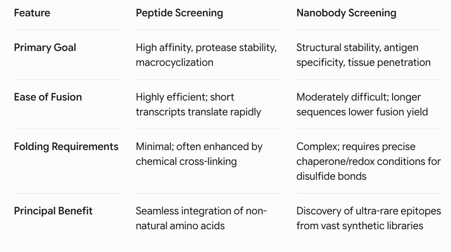
Tailoring mRNA Display for Peptides and Nanobodies
Conclusion
mRNA display is a pinnacle technology in the directed evolution landscape. For peptide discovery, it is virtually unrivaled, allowing scientists to explore chemical space beyond the 20 standard amino acids and discover robust macrocyclic drugs. For nanobodies, it provides an unparalleled library depth that can uncover rare therapeutic candidates that smaller phage libraries might miss—provided researchers can navigate the strict folding and synthesis requirements.
As cell-free protein synthesis kits become more sophisticated and automation simplifies the multi-step ligation process, mRNA display is poised to become an increasingly accessible and mainstream tool, reshaping the future of molecular biotherapeutics.